
CRANIOPLASTY

Patient-specific cranial plates are mainly used for reconstructive surgery after decompressive craniotomy after trauma, to shield the unprotected brain from external impact, pressure, and infections. When grafting the original bone flap or autologous split calvarial grafts are good options. Rather, a customized PEEK or Titanium cranial plate forms a fantastic alternative to acryl or Hydroxyapatite. However, patient-specific onlay cranioplasty can also be indicated to correct cranial deformations for aesthetic reasons. 3D printing together with traditional techniques allows us to integrate several state-of-the-art features. This results in reduced operating time and perfect aesthetic outcomes.
Product specifications
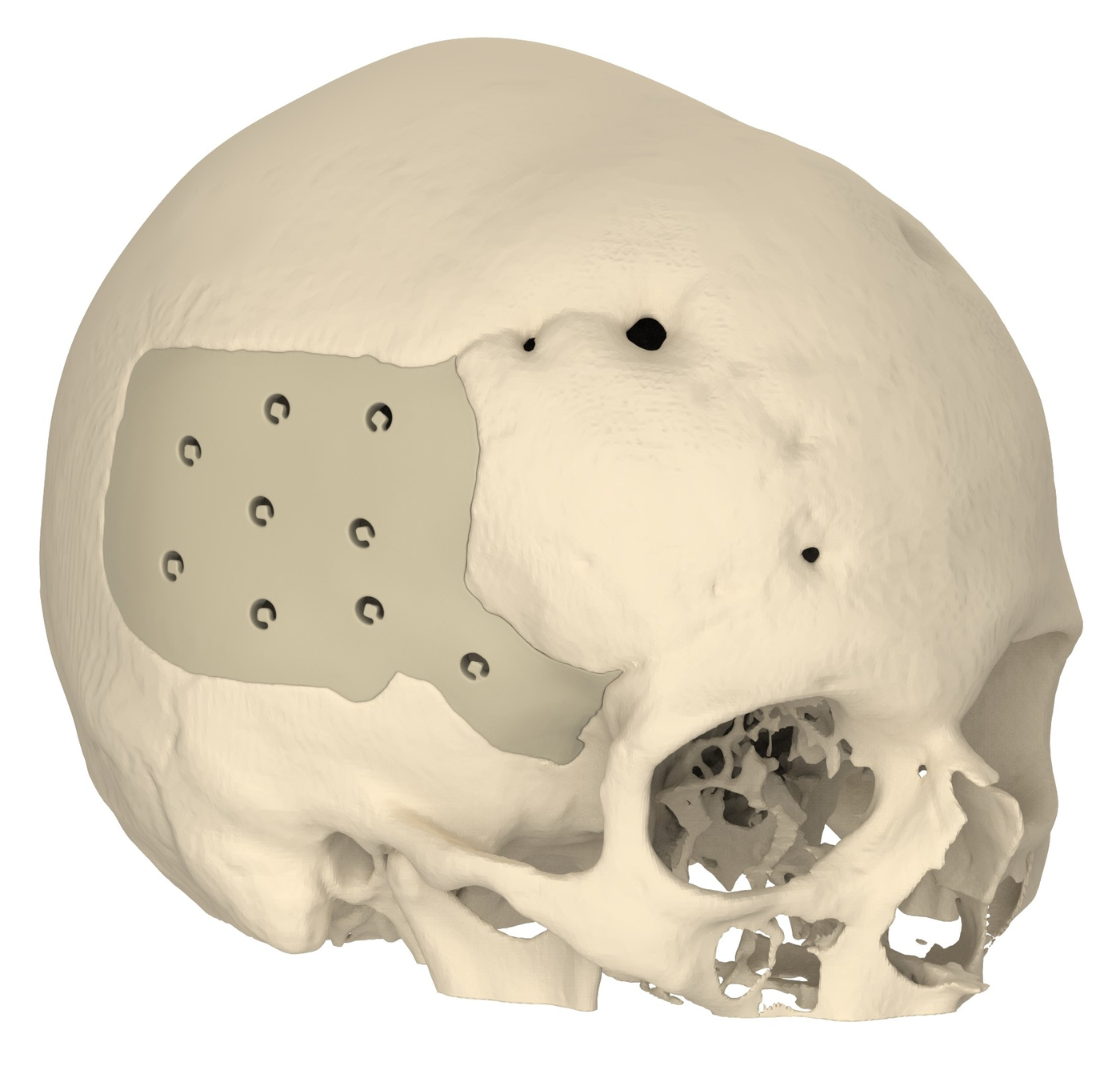
The design of the cranioplasty is done by mirroring the unaffected side to provide a symmetrical result. For large defects, the plate can be manufactured in multiple pieces, connected with a 3D-puzzle structure, and under-contoured for tensionless wound closure.
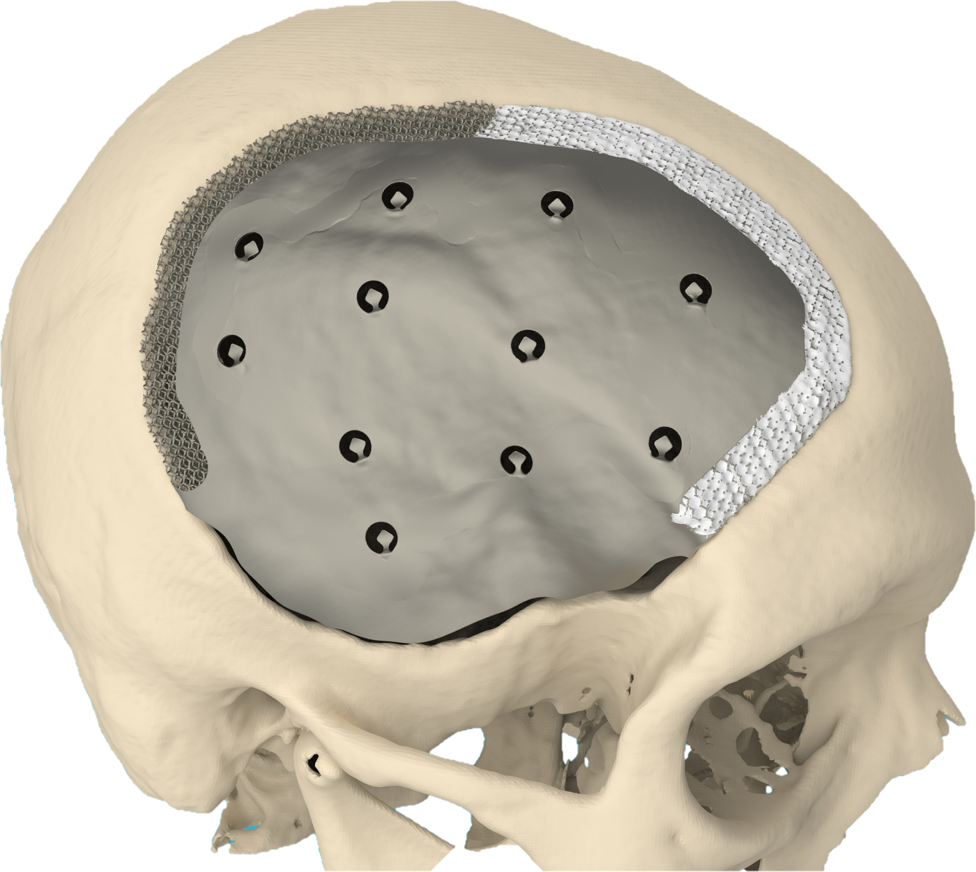
Perforations in the implants facilitate the drainage of cerebrospinal fluid and blood. Arrow-shaped brackets in the perforations serve as suspension fixation points for dura mater and temporalis muscle. To anticipate temporal hollowing following atrophy of the temporalis muscle, an extra onlay piece can be made to restore facial symmetry.
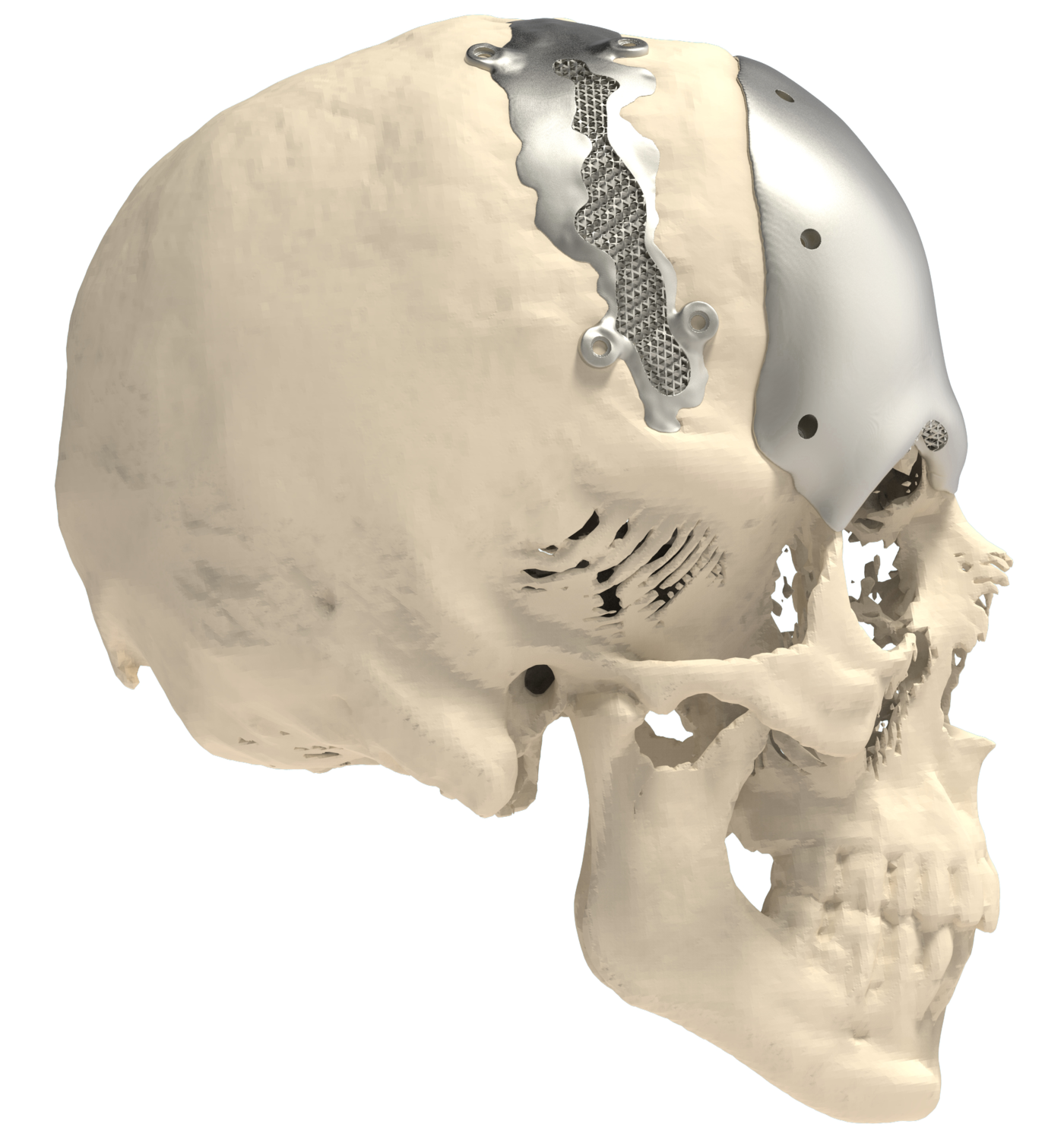
Our latest realization in cranioplasty is the CeTi. This stands for a combination of ceramics and titanium, combining the advantages of both materials. The CeTi is a titanium cranial plate with scaffolding at the border to include injectable hydroxyapatite bone substitute for optimal biocompatibility (e.g. Hydroset). The thin central part of the plate creates extra space if the brain expands and the specific design will absorb impact by deformation, evenly distributing the forces across the contact area, avoiding high local stresses on the skull.
CASE REPORT This patient receiving a Ceramic-Titanium (CeTi) cranioplasty had a history of a subarachnoid hemorrhage F4W1, caused by rupture of a right middle cerebral artery aneurysm that was clipped earlier. Explantation of the “bone flap”…
View clinical case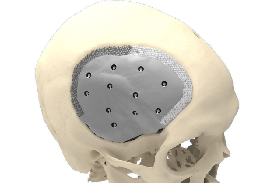
Contact us for more information,
fill in the form on the right or order
directly from your surgeon account.
Already have a surgeon account?