
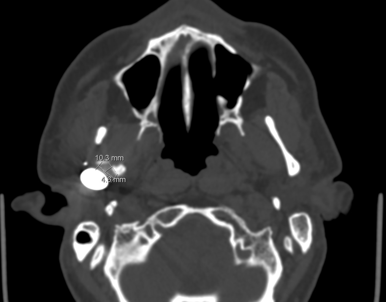
Evidence of enthesis reconstruction between lateral pterygoid muscle and lattice structure in pterygoid fovea of our CADskills 3D printed titanium mandibular component, visualized in axial CT scan.
3 months postoperative laterotrusion of 9 mm towards the healthy side, 10 mm towards the TJR. Mineralization of the callus between the osteotomized boney-tendon fragment and the bone chips in the lattice is evident. The indication was arthrosis, pain and recurrent condylar luxations after unrepaired subcondylar 25 years ago; not trismus. There was no ankylosis. Targetted ossification for enthesis reconstruction and reduction of unwonted calcifications are mandatory when cutting & grinding (protraction and lateral movement, hence normal mastication) are extra goals, besides achieving a normal mouth opening and reduction of pain.